Cervical Cancer Awareness Month: The Power of Imaging in Cervical Cancer
January 2024 – Imaging Endpoints (IE) is a global leader specializing in comprehensive imaging services for clinical trials. Our profound expertise includes analysis of CT, MRI, and PET/SPECT molecular imaging, particularly in the context of gynecologic malignancies, including cervical cancer.
Cervical cancer ranks as the fourth most prevalent cancer in women worldwide, as per the World Health Organization (WHO), with an estimated 604,000 new cases and 342,000 deaths reported in 2020. Squamous cell carcinoma (SCC) of the cervix constitutes 85% of all cervical cancers, while non-squamous cervical carcinomas, though less frequent (15%), carry a graver prognosis. The primary risk factor for cervical cancer is human papillomavirus (HPV) infection, alongside contributing factors such as immunosuppression and smoking.
The International Federation of Gynecology and Obstetrics (FIGO) clinical classification stands as the most widely used staging system for uterine cervical cancer. In 2018, FIGO recommended the incorporation of cross-sectional imaging (CT, MRI, PET-CT) into staging guidelines, as it offers a more accurate assessment of local disease and extrauterine spread. The precision of MR imaging in evaluating cervical tumor morphology, local extent, and its ability to assess factors like size, endocervical growth, uterine wall infiltration, and involvement of pelvic sidewall or adjacent organs (e.g., bladder, rectum) has significantly enhanced clinical staging and identification of prognostic factors. This advancement has led to improved and more suitable treatment options for women affected by this disease.
For women exhibiting clinical signs of tumor recurrence, molecular imaging with 18F-Fluorodexoyglucose (FDG) is the preferred modality, offering a comprehensive view of disease spread throughout the entire body. Recent innovations in functional imaging, including dynamic contrast-enhanced (DCE) MRI, diffusion-weighted imaging (DWI), texture imaging analysis, PET/MRI, and the use of various new radiotracers for PET imaging, have provided new excitement in visualizing and quantifying functional and structural aspects of cervical tumor characteristics. These aspects are closely associated with clinical outcomes, FIGO stage, lymph node metastases, prognostic histological tumor markers, treatment response, and, ultimately, patient outcome.
In summary, both conventional and functional imaging emerge as crucial non-invasive tools. Leveraging our extensive experience and expertise in standard and novel imaging technologies specific to these types of cancers, Imaging Endpoints stands at the forefront, ready to support your company’s imaging needs as we strive to bring potentially life-saving treatments to patients facing this formidable cancer.
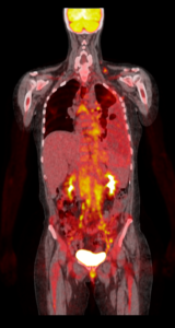
FDG PET/CT Scan in advanced Cervical Cancer
To learn how Imaging Endpoints can help your clinical trial succeed and aide in the war against Cervical Cancer,